My Blogging History

Saturday, January 10, 2015

WHAT YOU NEED TO KNOW ABOUT SCIATICA


The term sciatica describes the symptoms of leg pain and possibly tingling, numbness or weakness that originates in the lower back and travels through the buttock and down the large sciatic nerve in the back of the leg.  Sciatica (pronounced sighatihkah) is not a medical diagnosis in and of itself it is a symptom of an underlying medical condition.

SCIATICA NERVE PAIN

Sciatica is often characterized by one or more of the following symptoms:
  • Constant pain in only one side of the buttock or leg (rarely can occur in both legs)
  • Pain that is worse when sitting
  • Burning or tingling down the leg (vs. a dull ache)
  • Weakness, numbness or difficulty moving the leg or foot
  • A sharp pain that may make it difficult to stand up or to walk
Sciatica nerve pain pattern of inflammation
Sciatica Nerve Inflammation Pattern

Sciatic pain can vary from infrequent and irritating to constant and incapacitating. Specific sciatica symptoms also vary widely in type, location and severity, depending upon the condition causing the sciatica.

While symptoms can be very painful, it is rare that permanent sciatic nerve damage (tissue damage) will result.

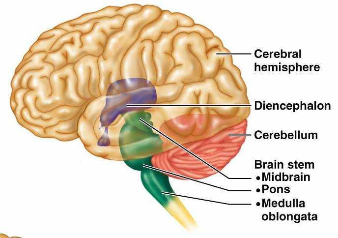
THE SCIATIC NERVE AND SCIATICA

The sciatic nerve is the largest single nerve in the body and is composed of individual nerve roots that start by branching out from the spine in the lower back and combine to form the "sciatic nerve." When it is irritated, sciatica symptoms occur.
  • The sciatic nerve starts in the lower back at lumbar segment 3 (L3).
  • At each level of the lower spine a nerve root exits from the inside of the spine and then comes together to make up the large sciatic nerve.
  • The sciatic nerve runs from the lower back, down the back of each leg
  • Portions of the sciatic nerve then branch out in each leg to innervate certain parts of the leg [e.g. the buttock, thigh, calf, foot, toes]
The sciatica symptoms (e.g., leg pain, numbness, tingling, weakness, possibly symptoms that radiate into the foot) are different depending on where the nerve is pinched. For example, a lumbar segment 5 (L5) nerve impingement can cause weakness in extension of the big toe and potentially in the ankle.

THE COURSE OF SCIATICA PAIN

The incidence of sciatica increases in middle age. Rarely occurring before age 20, the probability of experiencing sciatic pain peaks in the 50's and then declines.

Often, a particular event or injury does not cause sciatica, but rather it tends to develop over time. The vast majority of people who experience sciatica get better within a few weeks or months and find painrelief with non-surgical sciatica treatment. For others, however, sciatica pain from a pinched nerve can be severe and debilitating.

There are a few symptoms that may require immediate medical, and possibly surgical, intervention, such as progressive neurological symptoms (e.g. leg weakness) and/or bowel or bladder dysfunction (Cauda Equina Syndrome).

Because sciatica is caused by an underlying medical condition, treatment is focused on relieving the underlying causes of symptoms. Treatment is usually self-care and/or non-surgical, but for severe or intractable cases surgery may be an option.

MOST COMMON CAUSES OF SCIATICA

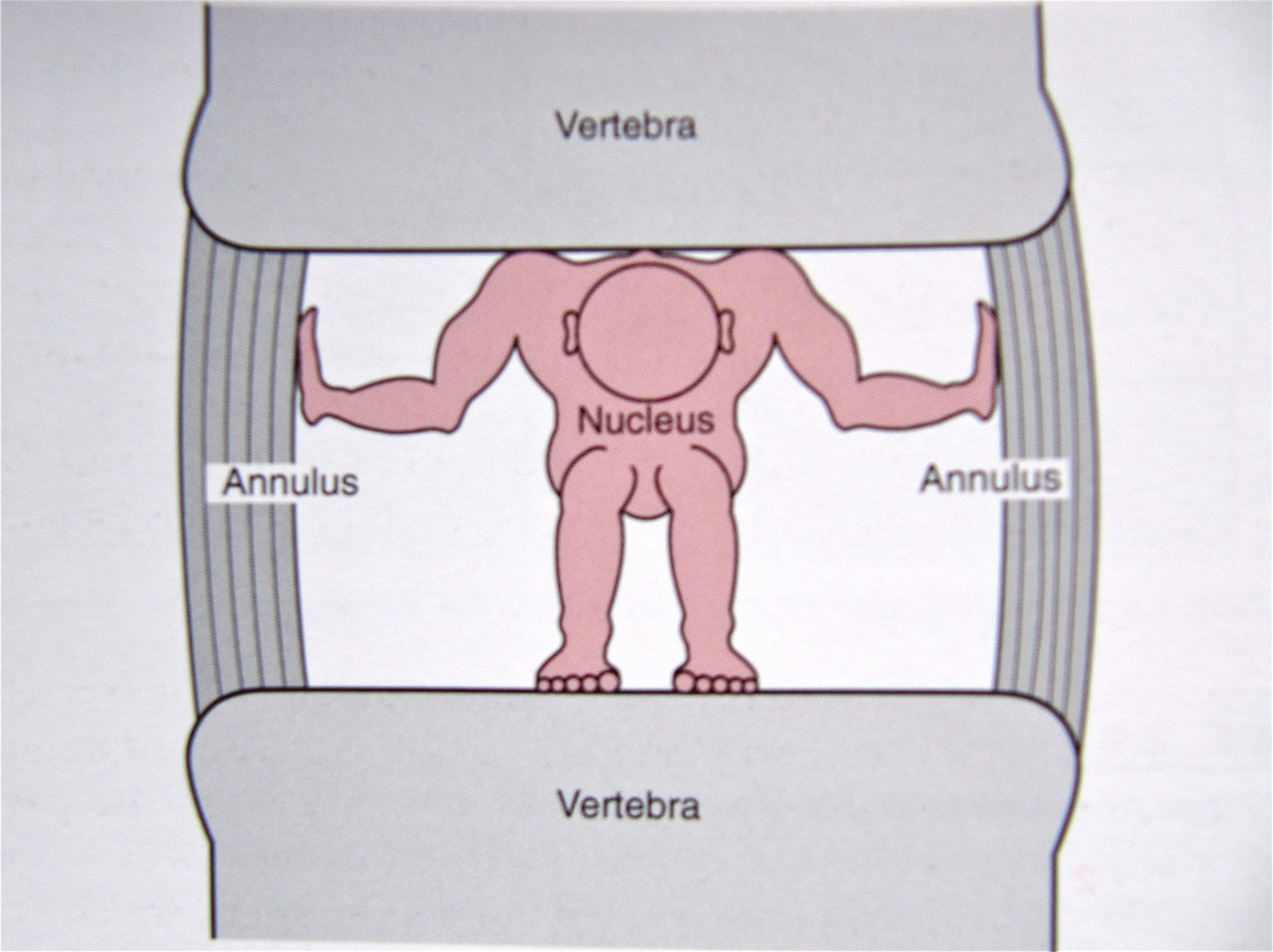
  • Lumbar herniated disc (also referred to as a slipped, ruptured, bulging, or protruding disc, or a pinched nerve) occurs when the soft inner core of the disc leaks out through the outer core and irritates the nerve root. Sciatica is the most common symptom of a lumbar herniated disc.

  • Degenerative disc disease is diagnosed when a weakened disc results in excessive micro-motion at that spinal level, and inflammatory proteins from inside the disc become exposed and irritate the area (including the nerve roots).

  • Isthmic spondylolisthesis. This condition occurs when a small stress fracture allows one vertebral body to slip forward on another (e.g. the L5 vertebra slips over the S1 vertebra). The combination of disc space collapse, the fracture, and the vertebral body slipping forward, can cause the nerve to get pinched and cause sciatica.

  • Lumbar spinal stenosis is related to natural aging in the spine and is relatively common in adults over age 60. The condition typically results from a combination of one or more of the following: enlarged facet joints, overgrowth of soft tissue, and a bulging disc placing pressure on the nerve roots, causing sciatica pain.

  • Piriformis syndrome. The sciatic nerve can get irritated as it runs under the piriformis muscle in the buttock. If the piriformis muscle irritates or pinches a nerve root that comprises the sciatic nerve, it can cause sciatica- type pain.

  • Sacroiliac joint dysfunction. Irritation of the sacroiliac joint - located at the bottom of the spine - can also irritate the L5 nerve, which lies on top of the sacroiliac joint, causing sciatica- type pain.


Monday, December 29, 2014

HOW A DISC BECOMES PAINFUL

This post was taken from our 18 July 14 Spine-Health Powered Newsletter


Each spinal disc is a unique and well-designed structure in the spine. It is strong enough to resist terrific forces in multiple different planes of motion, yet it is still highly mobile and permits motion in multiple directions.
The disc has several functions, including acting as a shock absorber between the bony vertebral bodies.

DISC ANATOMY AND FUNCTION

The intervertebral disc has been likened to a jelly donut. It is comprised of a series of bands that form a tough outer layer, and soft, jelly-like material contained within.
    - Annulus Fibrosus - the disc's firm, tough outer layer

Nerves to the disc space only penetrate into the very outer portion of the annulus fibrosus. Even though there is little innervation to the disc, it can become a significant source of back pain if a tear in the annulus reaches the outer portion and the nerves become sensitized.

With continued degeneration, the nerves on the periphery of the disc will actually grow further into the disc space and become a source of pain.
    - Nucleus Pulposus - the jelly-like inner disc material

The inner material contained in the disc, the nucleus pulposus, contains a great deal of inflammatory proteins. If this inner disc material leaks out of the disc and comes in contact with a nerve root, it will inflame the nerve root and create pain down the leg (sciatica or lumbar radiculopathy) or down the arm (cervical radiculopathy).

When we are born, the disc is comprised of about 80% water, which gives it its spongy quality and allows it to function as a shock absorber. As we age, the water content decreases and the disc becomes less capable of acting as a shock absorber

In the same manner, if any of the inflammatory proteins within the disc space leak out to the outer annulus and touch the pain fibers in this area, it can create a lot of low back pain or neck pain. (See Figure 1)

The proteins within the disc space also change composition, and most of us will develop tears into the annulus fibrosus (the outer hard core of the disc).
Most people will have some level of disc degeneration by their sixth decade, yet most do not have back pain (see Figure 2).

Degenerated Disc on MRI Scan

Magnetic Resonance Imaging, called an MRI scan, has contributed a great deal to our understanding of degenerative disc disease and the natural degenerative process. With the advent of MRI technology, good anatomic detail of the disc can be imaged and correlated with the individual's pain.

Through studies with MRI scans, it was found that:

  • A large number of young patients with chronic low back pain had evidence of disc degeneration on their MRI scans, and;
  • Up to 30% of young healthy adults with no back pain had disc degeneration on their MRI scans.

Variability in Degenerative Disc Disease

It is not exactly clear why some degenerated discs are painful and some are not.

There is probably a variety of reasons that discs can become painful.

Some theories about pain from degenerative disc disease are:
  • If a disc is injured or degenerated, it may become painful because of the resultant instability from the disc injury, which in turn can lead to an inflammatory reaction and causes low back pain.
  • Some people seem to have nerve endings that penetrate more deeply into the outer annulus than others, and this is thought to make the degenerated disc more susceptible to becoming a source of pain.

 While the exact causes are not known, there is a generally agreed upon theory of how a disc degenerates over time,....... Click here to read the full article: http://www.spine-health.com/conditions/degenerative-disc-disease/how-disc-becomes-painful


Tuesday, December 16, 2014

CHIROPRACTIC MANIPULATION FOR THE CERVICAL SPINE

This post is taken from our 11 July 14 Spine-Health Powered Newsletter


Manipulation of the cervical spine or neck region is a common technique utilized by doctors of chiropractic for many patients complaining of neck, upper back, and shoulder/arm pain, as well as headaches. Read more about this first line of treatment for cervical spine conditions.


TYPES OF CHIROPRACTIC MANIPULATION


The goals of cervical adjustment include reduction of pain, improved motion, and restored function. There are two general approaches for cervical spine complaints:

- Manipulation - often thought of as the traditional chiropractic adjustment, or a high-velocity, low-amplitude (HVLA) technique

- Mobilization - which is a more gentle/less forceful adjustment, or a low-velocity, low-amplitude (LVLA) technique moving the joint through a tolerable range of motion.

A combination of the various approaches varies among patients depending on the chiropractor's preferred techniques, the patient's comfort and preferences, and the patient's response to the treatment, as well as both past experience and observations made during the course of treatment.

Adjunctive therapies may include massage, therapeutic heat and/or cold application, gentle stretching and strengthening exercises, and more.


THE “CRACKING” SOUND

The HVLA manipulation usually results in a release, called cavitation, which is created in part by gas escaping from the joint capsule when the joint is moved quickly within its passive range of motion, well within the tissue boundaries.

This type of chiropractic adjustment creates the typical cracking sound that is often associated with joint manipulation. It sounds similar to cracking one's knuckles.

While this cracking description of a chiropractic high-velocity, low- amplitude thrust may give an impression of something that is uncomfortable, many patients find the sensation is relieving and may provide immediate relief of painful symptoms.

GENTLE MOBILIZATION OR ADJUSTMENT?

There are several reasons a chiropractor may recommend gentle (LVLA) chiropractic techniques, such as: 

- Patient preference: Some patients do not feel comfortable with the traditional high-velocity thrust manipulation and prefer an approach that does not involve twisting their neck or joint "popping."

- Provider experience: Some chiropractors may favor one technique and most chiropractors utilize several approaches and often modify and adapt several techniques to the patient’s needs and preferences.

- Contraindications: Some patients may not be able to tolerate the traditional chiropractic adjustment based on past experience or a prior injury or condition.


GENTLE TECHNIQUES
If the patient cannot relax, or simply dislikes the cracking feeling or sound, a gentle form of chiropractic adjustment may be utilized. Some of these methods include a slower technique performed within the passive range of joint motion. 

  • Cervical mobilization. The chiropractor manually moves the vertebra in the neck left to right, and vice versa, alternating between the side to side motion and a figure 8 movement, applied at varying degrees of moving the head forward, backward, and to the side and in rotation. This is a smooth, non-thrust type of stretch. 
  • Cervical manual traction. The chiropractor gently pulls on the neck, stretching the cervical spine often varying the angle between flexion (forward) and extension (backward), based on comfort and searching for the correct angle to most efficiently reduce the tightness. 
  • Cervical "drop" techniques. The chiropractor places a hand and/or finger over the spinal segment requiring the adjustment. A medium to high velocity, low amplitude thrust, or a non-thrust gradual increasing downward pressure is applied until the drop section / head piece of the table releases and drops a short distance. The goal is to reduce the fixation or restricted motion of the cervical vertebra assisted by the special table. 

TYPICAL CERVICAL PROBLEMS 

  • Mechanical neck pain includes pain generated from muscle, tendons, joint capsules, ligaments and/or the fascia. This type of problem is a common cause of neck pain and stiffness. Most of the time, many of these tissues are simultaneously injured and can cause pain both locally as well as at a distance away from the injury site. 
  • Cervical disc problems. Tears can develop in the cervical disc and/or the inside of the disc (the nucleus) can herniate through the outer part (the annulus) and trap or pinch the nerve root as it exits the spine. 
Cervical nerve root irritation can frequently refer pain down the arm and into the hand, usually affecting specific regions such as the 4th and 5th digits, the palm side thumb to 3rd fingers and/or the back of the hand on the thumb, index finger side of the hand, depending on which nerve root is irritated.

The above are two broad examples of types of cervical spine problems that may be treated with cervical spinal manipulation. The patient needs to receive a complete exam prior to cervical manipulation.


Click here to read the full article: http://www.spine-health.com/ treatment/chiropractic/chiropractic- manipulation-cervical-spine

Friday, October 17, 2014

“Conservative Care First” is more important today than ever in facing our nation’s healthcare challenges

Chiropractic physicians are the highest-rated healthcare practitioners for low-back pain treatments with their patient-centered, whole-person approach that provides greater interaction and communication for appropriate diagnosis and developing more cost-effective treatment planning.


October is National Chiropractic Health Month


Wednesday, September 17, 2014

What Everyone Should Know About Chiropractic


Friday, June 6, 2014

The Importance of Good Posture in Reducing Back Pain

The spine has three primary functions:
          1. It allows for movement between the different parts of the body
          2. It bears the forces applied to it (i.e. gravity, backpack, carrying a child, posture)
          3. It protects the spinal cord and exiting nerve roots

The spine requires muscle force and ligament tension operating within a control system so that it can resist buckling upon application of force (i.e. stretch, compression, shear, torsion) and subsequently maintain equilibrium[1].[2] This task is carried out by three distinct, yet intertwined, parts:
1. Vertebrae, spinal joints and their capsules, intervertebral discs, and spinal ligaments make up the Passive Musculoskeletal System. These components produce information regarding spinal position, the load(s) placed on each vertebra, as well as the motion (or lack thereof) of each vertebra, and via transducers[3] provide the Neural and Feedback System this information in real time.
Learn more about Spinal Anatomy <--- HERE
Learn more about Vertebral Discs  <--- HERE
Learn more about Spinal Ligaments <--- HERE

2. The Active Musculoskeletal System produces the forces necessary to carry out activities of daily living while simultaneously providing the spine its required stability. This system consists of the muscles and tendons that surround the spine.

3. The Neural and Feedback System determines the exact necessities for spinal stability via transducers located in every ligament, tendon, muscle, and neural control center (spinal cord gray matter, brainstem, cerebral cortex, cerebellum, and basal ganglia) that measure the forces and motions induced in/on the body. This sub-system then uses that information to determine the appropriate individual muscle tensions within the active sub-system so that it can fulfill its part of the stability mission. 

When functioning normally these three systems work interdependently[4] with moment to moment awareness of the demands placed upon the spine (posture as well as fixed and ambulatory[5] loads) to provide it with homeostatic  biomechanical [6] stability.

The average person is now asking him/herself, “Why is this information important to me?” Well this is the scientific reasoning behind why our mothers and grandmothers were continually harping about our need to “Sit/stand up straight”. In-vitro[7] experiments conducted at Yale and UC San Francisco found that vertebral segments in the thoracic and lumbar spine(s) became mechanically unstable at loads of 4.5 pound-force (20N) and 20.2 pound-force (90N) respectively.[8] [9] The irony of this is that the normal everyday loads placed on the spine from body mass alone while standing are significantly larger (20-times greater) and even more so when carrying a backpack, purse, groceries, etc. The only reason these segments do not buckle under these forces is because of the musculature surrounding the spine and why mindful adherence to good ergonomics/posture is imperative in preventing back and neck injury/pain. Over time, the stress of poor posture can change the anatomical characteristics of the spine, leading to the possibility of constricted blood vessels and nerves, as well as problems with muscles, discs, and joints.[10]

Envision a standard broom, better yet find a broom and with one hand grip the end of the handle and lift the broom in the air until it’s vertical. You will find a mechanically advantageous position once the broom is perpendicular to the floor; at this point holding the broom aloft is almost effortless. Anatomically this is considered “neutral position” a spinal posture in which the overall internal stresses in the spinal column and the muscular effort to hold the posture is minimal.[11] Now tip the broom slightly in any direction and experience how heavy it becomes, how much stress it now places on the wrist and arm, and how difficult it becomes to hold aloft with one hand.[12] This is equivalent to slouching, slumping, cradling a phone between your ear and shoulder, looking downward too often (text-neck); habits that over time expand the spine’s “neutral zone” and are the precedent to back and neck pain, headaches, fatigue, and over time respiratory and major organ dysfunction due to decreased stature. In fact one study showed that height loss greater than or equal to 1.18 inches (3cm) “in older men is independently associated with an increased risk of all-cause
mortality and coronary heart disease”.[13]

The spine’s “neutral zone” is the inner region of a joint’s range of motion where minimal resistance to motion is encountered.[2]To experience this directly take your thumb and first finger and grip the middle knuckle of one of the fingers on your opposite hand. Relax the gripped finger and begin to ever so slightly wiggle the finger back and forth. The slight motion you feel within that knuckle is the “neutral zone” which, as you've seen in this experiment, is restricted by the ligaments alone due to your voluntary removal of the active muscle forces that truly stabilize the joint. Poor posture and/or ergonomics stretch the spine’s passive subsystem (spinal joint capsules, intervertebral discs, and spinal ligaments) beyond its elastic limits and over time induces microtrauma which results in expansion of the “neutral zone” altering the signals sent by the transducers to the neural/feedback system. This places increased demands on the surrounding musculature degrading motor control. Consequentially inappropriate muscle activation sequences occur during simple tasks (i.e. bending over to pick up a piece of paper) transferring the force of these actions to the vertebral disc.
The center of the vertebral disc contains the nucleus pulpous which is a gelatinous cushion that protects the vertebrae from the pressures applied to the spine. This cushion is surrounded by several layers of fibrocartilage known as the annulus fibrosis which resemble the rings seen inside the trunk of a tree. Unsustainable pressures applied to the disc over time due to poor posture/ergonomics will create tearing in these rings which results in disc bulging and eventually herniation if one’s body mechanics/behaviors are not corrected.

Articles and Videos to Enhance the Importance of Good Posture/Body Mechanics


This information is not intended as a substitute for professional medical help or advice but is to be used only as an aid in understanding back pain.


[1] The ability to maintain the body’s center of mass over a stable base of support

[2] Liebenson, Craig. Rehabilitation of the Spine – A Practitioner’s Manual. 2nd ed. Baltimore: Lippincott Williams& Wilkins, 2007. Print

[3] A biological entity that converts energy in one form to another, e.g. the rods and cones of the eye or the hair cells of the ear

[4] Relying on mutual assistance, support, cooperation, or interaction among constituent parts

[5] Walking or moving around, or done while walking or moving

[6] The study of body movements and of the forces acting on the musculoskeletal system

[7] (Experimental Biology) Conducted using components of an organism (part of or a dead specimen)

[8] Crisco JJ. The biomechanical stability of the human lumbar spine: experimental and theoretical investigation [Doctoral Dissertation], New Haven, CT, Yale University, 1989

[9] Lucas DB, Bresler B. Stability of the ligamentous spine. Technical Report esr. 11 No. 40, Biomechanics Laboratory, University of California at San Francisco, The Laboratory

[10] Schubbe, John. "Good Posture Helps Reduce Back Pain." Spine-health: Trusted Information for Back Pain Relief. Spine-health.com, 17 May 2004. Web. 05 June 2014. <http://www.spine-health.com/wellness/ergonomics/good-posture-helps-reduce-back-pain>.

[11] Panjabi, Manohar M. "The Stabilizing System of the Spine. Part II. Neutral Zone and Instability Hypothesis." Journal of Spinal Disorders & Techniques 5.4 (1992): 390-96. Print.

[12] Weiniger, Steven P. Stand Taller ~ Live Longer: An Anti-Aging Strategy: 10 Minutes a Day to Keep Your Body Active and Pain-free. Alpharetta, GA: BodyZone, 2008. Print.

[13] S. Goya Wannamethee, PhD; A. Gerald Shaper, FRCP; Lucy Lennon, MSc; Peter H. Whincup, FRCP, PhD. "Height Loss in Older Men - Associations With Total Mortality and Incidence of Cardiovascular Disease." Arch Intern Med. 2006;166:2546-2552. Print.